What is Chordae Tendineae
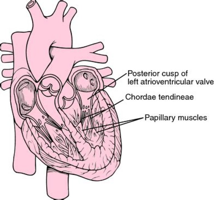
The chordae tendinae are the chord like structures connecting leaflets to the papillary muscle
What are chordae tendineae and what are their function?
Chordae tendineae: Thread-like bands of fibrous tissue which attach on one end to the edges of the tricuspid and mitral valves of the heart and on the other end to the papillary muscles, small muscles within the heart that serve to anchor the valves.
What is the chordae tendineae made of?
Chordae tendineae are composed of several types of ECM proteins, such as collagen type I, III, and fibronectin in the spongiosa layer, and collagen type III resides in the fibrosa layer [30].
What is the main function of chordae tendineae?
The chordae tendineae are a group of tough, tendinous strands in the heart. They are commonly referred to as the “heart strings” since they resemble small pieces of string. Functionally, the chordae tendineae play a vital role in holding the atrioventricular valves in place while the heart is pumping blood.What does Chordae mean?
chordae (kōr’dă, -dē) [TA] A tendinous or a cordlike structure. [L., cord]
What causes mitral valve to close?
The valve opens and closes because of pressure differences, opening when there is greater pressure in the left atrium than ventricle and closing when there is greater pressure in the left ventricle than atrium.
What is chordae tendineae in heart?
The chordae tendineae (tendinous cords), colloquially known as the heart strings, are inelastic cords of fibrous connective tissue that connect the papillary muscles to the tricuspid valve and the mitral valve in the heart.
What is the role of chordae tendineae and papillary muscles in the heart?
The bicuspid and tricuspid valves are connected to chordae tendineae which in turn are connected to the papillary muscles present on the ventricular wall. Chordae tendineae and papillary muscles regulate the opening and closing of valves.How do chordae tendineae support atrioventricular valves?
The two atrioventricular valves, the mitral valve, and the tricuspid valve, are connected to the ventricles by thin, fibrous strands of tissue called chordae tendineae. The chordae tendineae, along with papillary muscle hold the flaps, or cusps, of each valve in place.
Are the chordae tendineae elastic?In order to perform their function efficiently, the chordae have to possess a high degree of elasticity, as well as considerable strength and endurance. Human chordae tendineae originating from the left ventricles were obtained from 7 embalmed cadavers and 6 postmortem subjects of various ages.
Article first time published onWhat is a heartstring?
Definition of heartstring 1 obsolete : a nerve once believed to sustain the heart. 2 : the deepest emotions or affections —usually used in plural That movie really pulls at your heartstrings. Synonyms Heartstring Has a Medical History Example Sentences Learn More About heartstring.
How many chordae tendineae are there?
Five types of chordae were distinguished by their morphology and mode of insertion: Fan-shaped, rough zone, basal, free edge, and deep chordae.
What makes the heart valves open and close?
The heart valves open and close passively because of pressure differences on either side of the valve. When pressure is greater behind the valve, the leaflets are blown open and the blood flows through the valve. However, when pressure is greater in front of the valve, the leaflets snap shut and blood flow is stopped.
What is the function of papillary muscles?
Background— The papillary muscles (PMs) play an important role in normal cardiac function, helping to prevent leakage through the AV valves during systole. The nature of their attachment to the heart wall can affect the understanding of their function.
What happens if the chordae tendineae rupture?
Primary chordae tendineae rupture (CTR) can lead to a total loss of tension of one of the mitral valve leaflets, which then becomes flail. This often leads to abrupt aggravation of the MR, with fainting and/or acute congestive heart failure (CHF).
Which of the heart valves contain chordae tendineae?
The mitral valve is composed of two leaflets, the anterior (or aortic) and posterior leaflets. The supporting tendinous cords (chordae tendineae) on the ventricular aspect of the valve leaflets insert into two well-defined papillary muscles that are continuous with the left ventricular myocardium.
What is the moderator band?
In the human heart, the moderator band, or trabecula septomarginalis, is a muscle column that courses inferiorly from the right portion of the interventricular septum to the base of the anterior papillary muscle of the right ventricle This muscular structure is crossed by one or more arteries, which come from the …
Which valves of the heart have no chordae tendineae?
The semilunar valve on the left side of the heart is the aortic valve, named for the fact that it prevents the aorta from regurgitating blood back into the left ventricle. The semilunar valves are smaller than the AV valves and do not have chordae tendineae to Page 5 hold them in place.
How long can you live with leaky heart valve?
In developing countries, it progresses much more rapidly and may lead to symptoms in children less than 5 years of age. Around 80% of patients with mild symptoms live for at least 10 years after diagnosis.
What are the symptoms of a leaky mitral heart valve?
- Shortness of breath with exertion.
- Shortness of breath when lying flat.
- Tiredness (fatigue)
- Reduced ability to exercise.
- Unpleasant awareness of your heartbeat.
- Palpitations.
- Swelling in your legs, abdomen, and the veins in your neck.
- Chest pain (less common)
What are the symptoms of mitral valve disease?
- Fluttering or rapid heartbeat called palpitations.
- Shortness of breath, especially with exercise.
- Dizziness.
- Passing out or fainting , known as syncope.
- Panic and anxiety.
- Numbness or tingling in the hands and feet.
What are chordae tendineae in a fetal pig?
The chordae tendinae are thin strands of connective tissue that anchor the leaflets of each AV valve so that they cannot open into the atrium (thus allowing backflow of blood into the atrium).
What does chordae tendineae attach to?
The chordae tendineae are tendons linking the papillary muscles to the tricuspid valve in the right ventricle and the mitral valve in the left ventricle.
Why is it called atrioventricular valve?
The heart has two types of valves that keep the blood flowing in the correct direction. The valves between the atria and ventricles are called atrioventricular valves (also called cuspid valves), while those at the bases of the large vessels leaving the ventricles are called semilunar valves.
What occurs due to contraction of the papillary muscles quizlet?
What occurs due to contraction of the papillary muscles? The AV valves are prevented from protruding into the atria. … It allows an action potential to reach the left atrium so both atria contract together, before the ventricles contract.
What is the role of papillary muscle and tendon in human heart?
function in heart The papillary muscles project like nipples into the cavities of the ventricles. They are attached by fine strands of tendon to the valves between the atria and ventricles and prevent the valves from opening when the ventricles contract.
How do you spell Chordae Tendineae?
noun, plural chor·dae ten·din·e·ae [kawr-dee ten-din-ee-ee]. Anatomy. any of the tendons extending from the papillary muscles to the atrioventricular valves and preventing the valves from moving into the atria during ventricular contraction.
How many papillary muscles in the right and left ventricle?
Structure. There are five total papillary muscles in the heart; three in the right ventricle and two in the left. The anterior, posterior, and septal papillary muscles of the right ventricle each attach via chordae tendineae to the tricuspid valve.
What are the flaps on the front of the atria called?
These ear-like flaps are called auricles. 4. The front-most vessel is the pulmonary trunk.
What does a tricuspid valve do?
The tricuspid valve is on the right side of the heart. It separates the upper and lower chambers, also known as the right atrium and ventricle. The valve allows deoxygenated blood to flow through both of the chambers. The right ventricle pumps blood to the lungs, where it will be oxygenated.
Is Semilunar valve same as pulmonary valve?
cardiovascular system The semilunar valves are pocketlike structures attached at the point at which the pulmonary artery and the aorta leave the ventricles. The pulmonary valve guards the orifice between the right ventricle and the pulmonary artery.